Project ID: PI19/00458
Start Date: 25/11/2019
End Date: 01/01/2023
Funded Under: Instituto de Salud Carlos III
Overall Budget: € 15.600
Partners:
- Fundación Canaria de Investigación Sanitaria (FUNCANIS) – Proyect Coordinator
- Instituto Universitario de Microelectrónica Aplicada (IUMA) de la Universidad de Las Palmas de Gran Canaria (ULPGC)
Chemotherapy-induced peripheral neuropathy (CIPN) can cause severe pain, worsen other symptoms (anxiety, depression, insomnia, fatigue) and affect the ability to perform activities of daily living, decreasing the quality of life (QOL) of patients. It can also require a reduction in the dose of chemotherapy (CT) or even its interruption. Between 20% and 40% of cancer patients receiving neurotoxic CT may develop pain due to CIPN, 60% if platinum, taxanes, vincristine or bortezomib are used, and it could increase with the addition of antiangiogenic agents to new CT regimens. CIPN pain can persist for months or years after CT has finished, and prophylactic and therapeutic measures are very limited in number and degree of efficacy.
The marked discrepancy between the assessment of neurotoxicity made by doctors and that made by patients is noteworthy, which is why more relevance is currently being given to symptom assessment scales focused on the patients’ own assessment, with there being multiple scales subjective assessment. Furthermore, there are no valid objective diagnostic tests. Nerve conduction velocity tests are not necessarily related to pain and their value in clinical trials is not clearly established. They are performed together with an electromyogram, being an invasive test and involving a certain degree of discomfort.
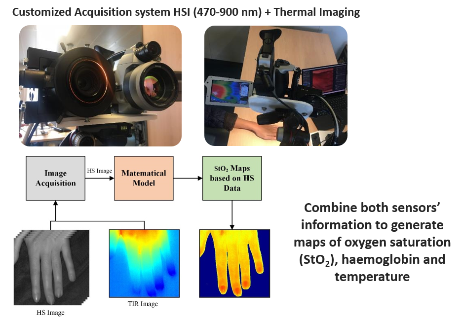
In this sense, thermography can detect small changes in skin temperature, related to physiological processes (flow, metabolism, function) and their alterations (ischemia, inflammation, dysregulation). It can be useful in the evaluation of neuropathic syndromes, such as complex regional pain syndrome, in the prediction of the appearance of post-herpetic neuropathy, and in the evaluation of neuropathic changes after ozone therapy. However, it does not seem to serve as an objective measure to assess the intensity of pain. Thermography analyzes tissue emission exclusively in the infrared range, while hyperspectral imaging studies analyze a much broader range of the spectrum, and not just infrared, which is why this technology is shown as an alternative non-invasive diagnosis potentially useful for this application.